Dysport vs. BOTOX® Cosmetic vs. Xeomin:
Which option is best for you?

Do you ever dread looking in the mirror? Are you tired of seeing your once youthful appearance succumb to the aging process? Time marches on, and unfortunately, those years of life experience can show more quickly on your face than other parts of your body, especially if you are a facially expressive person. That’s where injectables such as Botox, Dysport, and Xeomin may have crossed your mind. Botox vs. Dysport vs. Xeomin which is the best option?

Even with a proper skincare regime and skin maintenance, crow’s feet, forehead wrinkles, and frown lines are a natural part of life. The good news is, you don’t have to let your age show through your face. There are alternatives to plastic surgery that can provide you with smoother skin, fewer wrinkles, and a younger look. Injectables such as Botox, Xeomin, and Dysport, can help to soften the appearance of heavy wrinkles and lines and temporarily eliminate fine lines.

The trained professionals at Venice Avenue Dermatology are well versed in the benefits and administration of injectables and can recommend the best option for your specific skin needs. Call today to schedule your appointment and determine which neurotoxin treatment will work best for your skin needs.
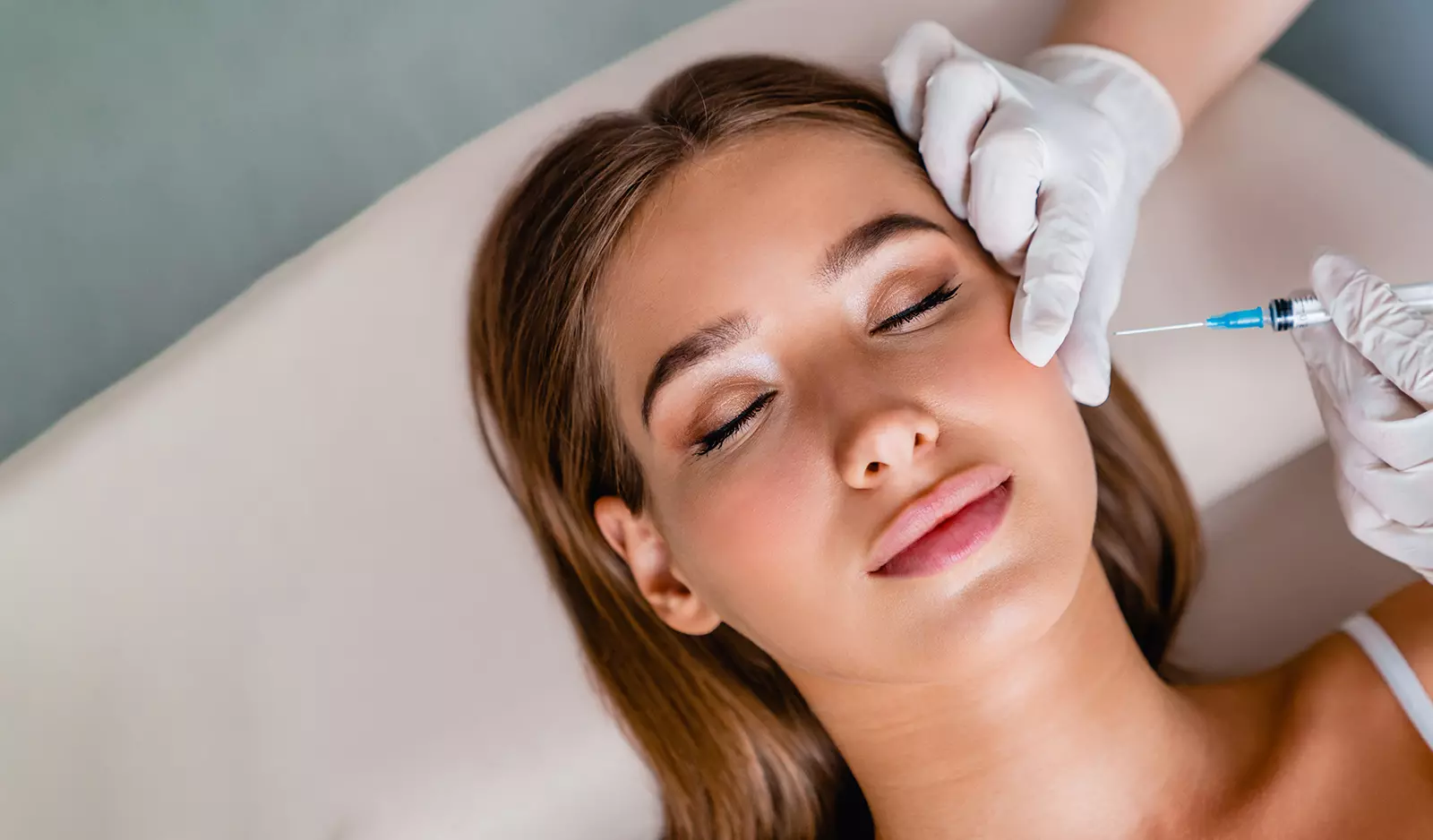
dysport vs botox

What are BOTOX® Cosmetic, Dysport, and Xeomin?

Botox Cosmetic, Dysport, and Xeomin are injectable neuromodulators that block nerve signals. When this occurs, the facial muscles responsible for most facial expressions are targeted, and muscle contractions are limited, reducing wrinkles. All three treatments fall under the category of non-invasive antiaging treatments.

All three are considered neurotoxins derived from botulinum toxin type A or bacterium Clostridium Botulinum. They are a popular anti-aging option as the treatment is quick with no downtime after the procedure. They are an excellent option for patients looking to reduce the appearance of frown lines, crow’s feet, and deep forehead wrinkles but don’t want to have a facelift.

What is the difference: Dysport vs. Botox vs. Xeomin?

While all three neurotoxins are similar in their make-up and function, they have slight differences. The formulation of Xeomin differs slightly from the others as it contains no additives and does not require refrigeration. They also vary slightly in the appearance of results. The effects of Botox spread three to five days after treatment when the full effects are seen. Xeomin can take even longer, with full results in roughly five to seven days. Dysport, or abobotulinumtoxina, is the option where results in the target areas will be seen most rapidly, with visible results within one to two days after treatment.

Xeomin and Dysport treatment also have greater diffusion than Botox, which allows them to reduce the appearance of wrinkles over a larger area. Botox is much more targeted and one of the most effective treatments for severe wrinkles in the forehead and glabellar lines.

Why choose a cosmetic dermatologist for neurotoxin injections like Botox, Xeomin, and Dysport?

Injectable neurotoxin treatments are aesthetic and can be administered at medical spas, by plastic surgeons, and in dermatology offices. While getting your anti-aging treatment at a medical spa may seem quick and easy, you will benefit more by having your procedure done at the dermatologist’s office.

Board-certified dermatologists and their staff care about the health and quality of your skin. They can help you discover the best options to achieve your desired results and address any underlying skin issues that could negatively affect your treatment. While considered a safe treatment option for fine lines and wrinkles, neurotoxins should be properly administered. Otherwise, you could end up with less than ideal results or more negative side effects.

By choosing a dermatologist for your injections, you will be given options for a more effective approach to your skin concerns and achieve better results from your treatment.

Which is better for crow’s feet: Dysport vs. Botox vs. Xeomin?

Botox is considered one of the best options for the treatment of crow’s feet. It has been the primary go-to for decades because it can tackle tough and deep facial lines and wrinkles. Full results may take longer to notice with Botox, but once the results have fully spread, you will see the best results for your crow’s feet with this option.

Which option is better for other fine lines?

Xeomin is one of the most popular choices for more fine facial lines. The product does not have preservatives and does not need to be refrigerated, making it more comfortable for injection and the more popular option for minor wrinkles. Xeomin requires fewer units for treating fine lines, making it an overall more cost-effective option for these types of lines. Xeomin is also ideal for those who have previously used Botox and Dysport and had resistance since it is considered a more purified form of neurotoxin.

Which option is better for forehead lines?

The fact that Botox is the most targeted option and works well on severe and deep wrinkles with fewer units makes Botox the best choice for horizontal forehead lines. The other two options can help to reduce forehead lines but may not soften them as significantly as Botox can.

What’s the difference between neurotoxin cosmetic injectables and dermal fillers?

Botox, Xeomin, and Dysport work by freezing facial muscles responsible for forming lines and wrinkles on the face. As long as these muscles are frozen, they cannot be used, which means your face will be more relaxed, leading to a softening of the facial profile. Dermal fillers address more static lines and hollow areas of the face by plumping up areas that have lost volume. This can help bring back fullness more common with a youthful appearance.

Neurotoxins and dermal fillers also differ in how they are administered. Neurotoxins are almost exclusively delivered into the upper part of the face, where wrinkles from facial muscles most often occur. Dermal fillers are injected anywhere on the face, especially around the lip and cheek area.

A final way the two non-invasive anti-aging treatments differ is in the length of time they last. Neurotoxins will last an average of three to four months, while dermal filler can last up to a year. Many patients who want to achieve an overall more youthful look to their face will often combine neurotoxin injectables and dermal filler treatments to tackle lines, facial wrinkles, and areas on their face that lack volume all at the same time.

Which lasts longer, BOTOX® Cosmetic or Dysport or Xeomin?

When properly administered by a certified injector, all three options should last between three and four months before additional injections are needed. Some patients have reported shorter time frames between injections, while others have been able to go the full three to four months between treatments. Some patients have reported that although the effects of Dysport tend to show up sooner, they fade slightly sooner as well.

dysport vs. botox

How many units of Dysport vs. BOTOX® Cosmetic vs. Xeomin will I need?

Dysport, Xeomin, and Botox injections require a specific number of units to reduce the appearance of wrinkles and achieve youthful results. Overall, Botox will require the least number of units. Deep forehead wrinkles and crow’s feet typically require 10 to 15 units to be effective, and frown lines can require between 20 and 30 units.

Xeomin comes in a close second, requiring between 15 and 30 units for frown lines and between 20 and 40 units to tackle tough forehead wrinkles. Dysport requires the most units, making it a more expensive option even though the units cost less. You will need between 30 to 50 units of Dysport for your crow’s feet and around 60 units for deep glabellar lines.

Are Botox, Xeomin, and Dysport the same price?

Some options are priced differently per unit, and each option requires a different number of units to achieve the desired result, which makes the overall cost of each treatment type different. Xeomin and Botox typically run in the same average range at $12 to $17 per unit. Still, Xeomin requires more units to complete treatment, in most cases, which can make Xeomin treatment more expensive than Botox. Dysport injections run an average of $4 to $6 per unit. While Dysport may cost less per unit than the other options, typically twice as many units of Dysport are required for most treatments.

Which is more natural looking?

All three products can reduce the signs of aging and be a natural-looking wrinkle treatment if appropriately done by a certified professional. Though out of the three, Xeomin is the more concentrated and pure form of Botulinum toxin as it contains no preservative proteins. It is sometimes referred to as“naked injectable.”

What are the side effects of Dysport vs. Botox vs. Xeomin compared?

All three products have been rigorously tested by the FDA and received FDA approval, meaning they are considered to be safe for most people if properly administered. Each product uses the same active ingredients, which makes the botox side effects similar each, though patients with certain medical conditions may have more side effects than others. The most common side effects associated with Dysport, Botox, and Xeomin include:

  • Eye dryness
  • Blurred vision
  • Droopy eyelids
  • Headache
  • Muscle spasms
  • Pain, bruising, or irritation at the injection site or treatment area

In most cases, side effects fade rather quickly.

How long does Dysport vs. Botox vs. Xeomin last?

All three products are designed to stay in effect for 3 to 4 months before additional injections are needed, though the actual length can vary from person to person.

Ready to take control of your skincare needs and find the right neurotoxin treatment to help you turn back the clock? The professionals at Venice Avenue Dermatology are here to help you fight the signs of aging and enjoy the more youthful look you desire. Schedule an appointment today to take the first step to start seeing a more youthful you in the mirror.

Skin Care Basics

The skin is the body’s largest organ and accounts for roughly 18% of an adult’s weight. It serves as a protective outer layer that keeps in moisture and keeps out invasive organism (like infections). It protects our organs against injury. It also helps regulate the body’s temperature and has self-healing capabilities.

The best way to maintain healthy skin is to prevent skin damage from occurring in the first place. Wrinkles, age spots and leathery patches are all the result of skin damage from overexposure to ultraviolet radiation from the sun. But the aging process for skin is unavoidable. As we age, skin becomes dryer and thinner. Repeated movements of facial muscles, such as frowning, smiling or squinting, cause wrinkles over time. Stress, gravity and obesity also contribute to aging skin. And because the skin is thinner, it is more susceptible to bruising.

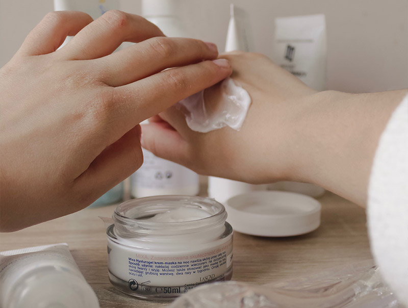
Person applying lotion to hand image

Photoaging

The premature aging of the skin from ultraviolet light exposure is called photoaging. Photoaging occurs when ultraviolet radiation penetrates deep into the dermis, damaging collagen fibers and causing the increased production of abnormal elastin. This breakdown in fundamental skin structures leads to deep wrinkles, fine lines, discoloration of the skin (age or liver spots), leatheriness and sagging skin.

Skin Care Routine

A healthy skin care routine throughout life can reduce the symptoms of aging in the skin. Be sure to:

  • Wash your face using a gentle cleanser and lukewarm water twice a day.
  • Pat skin dry; don’t rub it dry.
  • Exfoliate the skin twice a week to remove dead cells.
  • Apply a moisturizer to skin immediately after a shower or bath.
  • Wear sunscreen with a SPF of at least 15 every day.
  • For women who wear makeup, be sure to leave time each day when the skin is clean and free of makeup.
  • Do not use tanning beds.
  • Maintain a healthy diet and drink lots of water.
  • Get an adequate amount of sleep every day.
  • Quit smoking.
  • Avoid stress.
  • Conduct a monthly self-examination of your skin to detect any changes that might lead to cancer.
  • See your dermatologist once a year.

Anti-Aging Treatments

Beyond prevention, in today’s world there is a wide range of options for slowing down the affects of aging on the skin. See the Cosmetic Dermatology section of this website for more information about:

Skin Infections

Anyone who has a break in the skin is at risk for an infection. There are three types of skin infections:

Bacterial Infection

There are many bacteria that live on the surface of healthy skin. But with a break in the skin, these bacteria can invade the outer layer of skin and cause an infection and rash. Staph is a common cause of bacterial infections of the skin. Impetigo is one of the most common causes of skin infections in children. Oral or topical antibiotics are used to treat bacterial skin infections.

Viral Infection

Viruses are parasitic organisms that can live and grow inside living cells. They cause either a degeneration or a proliferation of the cell. Most causes of viral skin infections are either from Human Papilloma Virus, which causes warts, or Human Herpes Virus, which causes cold sores, chicken pox, shingles, genital herpes and mononucleosis. Viruses do not respond to antibiotics. Generally, medications are prescribed to help alleviate the symptoms of the infection, such as a rash or itch. Additionally, vaccinations are used to prevent viral infections.

Fungal Infections

Fungal infections of the human body are called mycoses and affect only the outer layer of skin. Although seen in all areas of the body, skin mycoses most frequently appear as yeast infections, thrush, athlete’s foot or jock itch.

Sun Safety

Because of the ultraviolet radiation it emits, the sun is inherently dangerous to human skin. In fact, the American Academy of Dermatology stipulates that there is no safe way to tan. Tanning is the skin’s natural response to damage from the sun. Additionally, the Environmental Protection Agency proclaims that everybody, regardless of race or ethnicity, is subject to the potential adverse effects of overexposure to the sun. That’s why everyone needs to protect their skin from the sun every day.

How We Burn

When ultraviolet light penetrates the epidermis it stimulates melanin, the substance responsible for skin pigmentation. Up to a point, the melanin absorbs dangerous UV rays before they do serious damage. Melanin increases in response to sun exposure, which is what causes the skin to tan. This is a sign of skin damage, not health. Sunburns develop when the UV exposure is greater than the skin’s natural ability to protect against it.

Child on beach wearing bucket hat image

Sunscreens and Sunblocks

The sun emits two types of ultraviolet (UV) rays that are harmful to human skin. UVA rays penetrate deep into the dermis and lead to wrinkles, age spots and skin cancers. UVB rays are responsible for causing sunburn, cataracts and immune system damage. Melanoma is thought to be associated with severe UVB sunburns that occur before the age of 20.

Sunscreens absorb ultraviolet light so that it doesn’t reach the skin. Look for sunscreens with the active ingredients PABA, benzophenones, cinnamates or salicylates. Sunblocks literally block the UV rays instead of absorbing them. Key active ingredients for sunblock success are titanium oxide and zinc oxide.

There is no sunscreen or sunblock that works 100%. The U.S. Food and Drug Administration regulates the manufacture and promotion of sunscreens. Sunscreens are given a SPF (Sun Protection Factor) number that indicates how long a person can remain in the sun without burning. It is recommended that people use products with a SPF of 15 or greater. Sunscreens are not generally recommended for infants six months old or younger. Infants should be kept in the shade as much as possible and should be dressed in protective clothing to prevent any skin exposure and damage.

There is no such thing as “all-day protection” or “waterproof” sunscreen. No matter what the SPF number, sunscreens need to be re-applied every 2 to 3 hours. Products that claim to be “waterproof” can only protect against sunburn up to 80 minutes in the water. Products labeled “water resistant” can only protect against sunburn up to 40 minutes in the water.

Even in the worst weather, 80% of the sun’s UV rays can pass through the clouds. Additionally, sand reflects 25% of the sun’s UV rays and snow reflects 80% of the sun’s UV rays. That’s why sunscreen needs to be worn every day and in every type of weather and climate. The sun’s intensity is also impacted by altitude (the higher the altitude the greater the sun exposure), time of year (summer months) and location (the closer to the Equator, the greater the sun exposure).

Protecting Yourself From Sun Exposure

  • Look for sunscreens that use the term “broad spectrum” because they protect against both UVA and UVB rays.
  • Choose a sunscreen with a minimum SPF rating of 15.
  • Apply sunscreen 15 to 30 minutes before you head out into the sun to give it time to seep into the skin.
  • Apply sunscreens liberally. Use at least one ounce to cover the entire body.
  • Use a lip balm with SPF 15 or greater to protect the lips from sun damage.
  • Re-apply sunscreen immediately after going into water or sweating.
  • Re-apply sunscreen every 2 to 3 hours.
  • Use sunscreen every day regardless of the weather.
  • Wear sunglasses to protect the eyes from UV rays.
  • Wear wide-brimmed hats and protective clothing to limit skin exposure to the sun.
  • Stay in the shade whenever possible.
  • Avoid using tanning beds.

Treating a Sunburn

If you experience a sunburn, get out of the sun and cover the exposed skin as soon as possible. A sunburn will begin to appear within 4 to 6 hours after getting out of the sun and will fully appear within 12 to 24 hours. Mild burns cause redness and some peeling after a few days. They can be treated with cold compresses on the damaged area, cool baths, moisturizers to prevent dryness and over-the-counter hydrocortisone creams to relieve any pain or itching. It is also important to drink plenty of fluids when you experience any type of sunburn.

More serious burns lead to blisters, which can be painful. It is important not to rupture blisters as this slows down the natural healing process and may lead to infection. You may want to cover blisters with gauze to keep them clean. Stay out of the sun until your skin has fully healed. In the most severe cases, oral steroids may be prescribed to prevent or eliminate infection along with pain-relieving medication.

Skin Cancers

Skin cancer is the most common form of human cancers, affecting more than one million Americans every year. One in five Americans will develop skin cancer at some point in their lives. Skin cancers are generally curable if caught early. However, people who have had skin cancer are at a higher risk of developing a new skin cancer, which is why regular self-examination and doctor visits are imperative.

The vast majority of skin cancers are composed of three different types: basal cell carcinoma, squamous cell carcinoma and melanoma.

Basal Cell Carcinoma

This is the most common form of skin cancer. Basal cells reside in the deepest layer of the epidermis, along with hair follicles and sweat ducts. When a person is overexposed to UVB radiation, it damages the body’s natural repair system, which causes basal cell carcinomas to grow. These tend to be slow-growing tumors and rarely metastasize (spread). Basal cell carcinomas can present in a number of different ways:

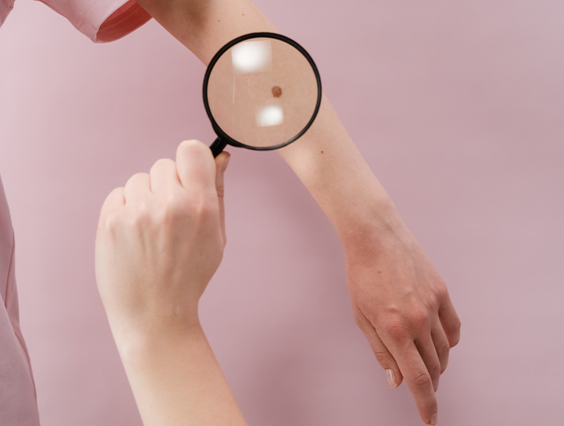
Cancerous mole on arm image
  • raised pink or pearly white bump with a pearly edge and small, visible blood vessels
  • pigmented bumps that look like moles with a pearly edge
  • a sore that continuously heals and re-opens
  • flat scaly scar with a waxy appearance and blurred edges

Despite the different appearances of the cancer, they all tend to bleed with little or no cause. Eighty-five percent of basal cell carcinomas occur on the face and neck since these are areas that are most exposed to the sun.

Risk factors for basal cell carcinoma include having fair skin, sun exposure, age (most skin cancers occur after age 50), exposure to ultraviolet radiation (as in tanning beds) and therapeutic radiation given to treat an unrelated health issue.

Diagnosing basal cell carcinoma requires a biopsy — either excisional, where the entire tumor is removed along with some of the surrounding tissue, or incisional, where only a part of the tumor is removed (used primarily for large lesions).

Treatments for basal cell carcinoma include:

  • Cryosurgery — Some basal cell carcinomas respond to cryosurgery, where liquid nitrogen is used to freeze off the tumor.
  • Curettage and Desiccation — The preferred method of dermatologists, this treatment involves using a small metal instrument (called a curette) to scrape out the tumor along with an application of an electric current into the tissue to kill off any remaining cancer cells.
  • Mohs Micrographic Surgery — The preferred method for large tumors, Mohs Micrographic Surgery combines removal of cancerous tissue with microscopic review while the surgery takes place. By mapping the diseased tissue layer by layer, less healthy skin is damaged when removing the tumor.
  • Prescription Medicated Creams — These creams can be applied at home. They stimulate the body’s natural immune system over the course of weeks.
  • Radiation Therapy — Radiation therapy is used for difficult-to-treat tumors, either because of their location, severity or persistence.
  • Surgical Excision — In this treatment the tumor is surgically removed and stitched up.

Squamous Cell Carcinoma

Squamous cells are found in the upper layer (the surface) of the epidermis. They look like fish scales under a microscope and present as a crusted or scaly patch of skin with an inflamed, red base. They are often tender to the touch. It is estimated that 250,000 new cases of squamous cell carcinoma are diagnosed annually, and that 2,500 of them result in death.

Squamous cell carcinoma can develop anywhere, including inside the mouth and on the genitalia. It most frequently appears on the scalp, face, ears and back of hands. Squamous cell carcinoma tends to develop among fair-skinned, middle-aged and elderly people who have a history of sun exposure. In some cases, it evolves from actinic keratoses, dry scaly lesions that can be flesh-colored, reddish-brown or yellow black, and which appear on skin that is rough or leathery. Actinic keratoses spots are considered to be precancerous.

Like basal cell carcinoma, squamous cell carcinoma is diagnosed via a biopsy — either excisional, where the entire tumor is removed along with some of the surrounding tissue, or incisional, where only a part of the tumor is removed (used primarily for large lesions).

Treatments for basal cell carcinoma include:

  • Cryosurgery Some basal cell carcinomas respond to cryosurgery, where liquid nitrogen is used to freeze off the tumor.
  • Curettage and Desiccation — The preferred method of dermatologists, this treatment involves using a small metal instrument (called a curette) to scrape out the tumor along with an application of an electric current into the tissue to kill off any remaining cancer cells.
  • Mohs Micrographic Surgery — The preferred method for large tumors, Mohs Micrographic Surgery combines removal of cancerous tissue with microscopic review while the surgery takes place. By mapping the diseased tissue layer by layer, less healthy skin is damaged when removing the tumor.
  • Prescription Medicated Creams — These creams can be applied at home. They stimulate the body’s natural immune system over the course of weeks.
  • Radiation Therapy — Radiation therapy is used for difficult-to-treat tumors, either because of their location, severity or persistence.
  • Surgical Excision — In this treatment the tumor is surgically removed and stitched up.

Melanoma

While melanoma is the least common type of skin cancer, it is by far the most virulent. It is the most common form of cancer among young adults age 25 to 29. Melanocytes are cells found in the bottom layer of the epidermis. These cells produce melanin, the substance responsible for skin pigmentation. That’s why melanomas often present as dark brown or black spots on the skin. Melanomas spread rapidly to internal organs and the lymph system, making them quite dangerous. Early detection is critical for curing this skin cancer.

Melanomas look like moles and often do grow inside existing moles. That’s why it is important for people to conduct regular self-examinations of their skin in order to detect any potential skin cancer early, when it is treatable. Most melanomas are caused by overexposure to the sun beginning in childhood. This cancer also runs in families.

Melanoma is diagnosed via a biopsy. Treatments include surgical removal, radiation therapy or chemotherapy.

What to Look For

The key to detecting skin cancers is to notice changes in your skin. Look for:

  • Large brown spots with darker speckles located anywhere on the body.
  • Dark lesions on the palms of the hands and soles of the feet, fingertips toes, mouth, nose or genitalia.
  • Translucent pearly and dome-shaped growths.
  • Existing moles that begin to grow, itch or bleed.
  • Brown or black streaks under the nails.
  • A sore that repeatedly heals and re-opens.
  • Clusters of slow-growing scaly lesions that are pink or red.

The American Academy of Dermatology has developed the following ABCDE guide for assessing whether or not a mole or other lesion may be becoming cancerous.

Asymmetry: Half the mole does not match the other half in size, shape or color.

Border: The edges of moles are irregular, scalloped, or poorly defined.

Color: The mole is not the same color throughout.

Diameter: The mole is usually greater than 6 millimeters when diagnosed, but may also be smaller.

Evolving: A mole or skin lesion that is different from the rest, or changes in size, shape, or color.

If any of these conditions occur, please make an appointment to see one of our dermatologists right away. The doctor may do a biopsy of the mole to determine if it is or isn’t cancerous.

Prevention

Roughly 90% of nonmelanoma cancers are attributable to ultraviolet radiation from the sun. That’s why prevention involves:

  • Staying out of the sun during peak hours (between 10 a.m. and 4 p.m.).
  • Covering up the arms and legs with protective clothing.
  • Wearing a wide-brimmed hat and sunglasses.
  • Using sunscreens year round with a SPF of 15 or greater and sunblocks that work on both UVA and UVB rays. Look for products that use the term “broad spectrum.”
  • Checking your skin monthly and contacting your dermatologist if you notice any changes.
  • Getting regular skin examinations. It is advised that adults over 40 get an annual exam with a dermatologist.

Skin Growths

Seborrheic Keratosis

Also known as seborrheic verruca, most people will develop at least one seborrheic keratosis during a lifetime. Fortunately, these lesions are benign and don’t become cancerous. They are characterized as brown, black or yellow growths that grow singly or in groups and are flat or slightly elevated. Often they are mistaken for warts. Generally, no treatment is required unless the growth becomes irritated from chafing against clothing. However, because it look similar in appearance to precancerous growths (actinic keratosis), your dermatologist will likely biopsy the tissue to confirm the diagnosis.

If a seborrheic keratosis becomes irritated or unsightly, removal is conducted using one of these three methods:

  • Cryosurgery, which freezes off the growth using liquid nitrogen.
  • Curettage, in which the doctor scrapes the growth off the surface of the skin.
  • Electrocautery, used alone or in conjunction with curettage to burn off the tissue and stop the bleeding.
Woman with facial mole image

Moles (Nevi)

Moles are brown or black growths, usually round or oval, that can appear anywhere on the skin. They can be rough or smooth, flat or raised, single or in multiples. They occur when cells that are responsible for skin pigmentation, known as melanocytes, grow in clusters instead of being spread out across the skin. Generally, moles are less than one-quarter inch in size. Most moles appear by the age of 20, although some moles may appear later in life. Most adults have between 10 and 40 moles. Because they last about 50 years, moles may disappear by themselves over time.

Most moles are harmless, but a change in size, shape, color or texture could be indicative of a cancerous growth. Moles that have a higher-than-average chance of becoming cancerous include:

Congenital Nevi

Moles present at birth. The larger their size, the greater the risk for developing into a skin cancer.

Atypical Dysplastic Nevi

Irregularly shaped moles that are larger than average. They often appear to have dark brown centers with light, uneven borders.

Higher frequency of moles

People with 50 or more moles are at a greater risk for developing a skin cancer.

In some cases, abnormal moles may become painful, itchy, scaly or bleed. It’s important to keep an eye on your moles so that you can catch any changes early. We recommend doing a visual check of your body monthly, including all areas that don’t have sun exposure (such as the scalp, armpits or bottoms of feet).

Use the American Academy of Dermatology’s ABCDEs as a guide for assessing whether or not a mole may be becoming cancerous:

Asymmetry: Half the mole does not match the other half in size, shape or color.

Border: The edges of moles are irregular, scalloped, or poorly defined.

Color: The mole is not the same color throughout.

Diameter: The mole is usually greater than 6 millimeters when diagnosed, but may also be smaller.

Evolving: A mole or skin lesion that is different from the rest, or changes in size, shape, or color.

If any of these conditions occur, please make an appointment to see one of our dermatologists right away. The doctor may do a biopsy of the mole to determine if it is or isn’t cancerous and/or may surgically remove it.

Warts

Warts are small, harmless growths that appear most frequently on the hands and feet. Sometimes they look flat and smooth, other times they have a dome-shaped or cauliflower-like appearance. Warts can be surrounded by skin that is either lighter or darker. Warts are caused by different forms of Human Papilloma Virus (HPV). They occur in people of all ages and can spread from person-to-person and from one part of the body to another. Warts are benign (noncancerous) and generally painless. They may disappear without any treatment. However, in most cases eliminating warts takes time.

The location of a wart often characterizes its type:

Common warts can appear anywhere on the body, although they most often appear on the back of fingers, toes and knees. These skin-colored, dome-shaped lesions usually grow where the skin has been broken, such as a scratch or bug bite. They can range in size from a pinhead to 10mm and may appear singly or in multiples.

Filiform warts look like a long, narrow, flesh-colored stalk that appears singly or in multiples around the eyelids, face, neck or lips. They are sometimes called facial warts. They may cause itching or bleeding, but are easy to treat with over-the-counter medications.

Flat (plane) warts appear on the face and forehead. They are flesh-colored or white, with a slightly raised, flat surface and they usually appear in multiples. Flat warts are more common among children and teens than adults.

Genital warts appear around the genital and pubic areas. It is also possible to get genital warts inside the vagina and anal canal or in the mouth (known as oral warts). The lesions start small and soft but can become quite large. They often grow in clusters. They are both sexually transmitted and highly contagious. In fact, it is recommended you generally avoid sex with anyone who has a visible genital wart. Genital warts should always be treated by a physician.

Plantar warts appear on the soles of the feet and can be painful since they are on weight-bearing surfaces. They have a rough, cauliflower-like appearance and may have a small black speck in them. They often appear in multiples and may combine into a larger wart called a mosaic wart. Plantar warts can spread rapidly.

Subungual and periungual warts appear as rough growths around the fingernails and/or toenails. They start as nearly undetectable, pin-sized lesions and grow to pea-sized with rough, irregular bumps with uneven borders. Subungual and periungual warts can impede healthy nail growth. Because of their location, they are difficult to treat and generally require medical attention.

Most warts respond to over-the-counter treatments, including:

  • Cryotherapy, which freezes off the wart using liquid nitrogen or nitrous oxide.
  • Electrosurgery, which sends an electric current through the wart to kill the tissue.
  • Laser surgery, which essentially heat up the wart until the tissue dies and the wart eventually falls off.
  • Nonprescription freezing products (dimethyl ether), aerosol sprays that freeze the warts and cause them to die off.
  • Salicylic acid preparations, which dissolve the protein (keratin) that makes up the wart and the thick layer of skin that covers it. It comes in gels, pads, drops and plasters and takes 4 to 6 weeks to eradicate the warts.

If self-treatments don’t work after a period of about 4 to 12 weeks, contact our dermatologist. We’ll assess your warts and recommend the best option.

Always contact the dermatologist if a wart is causing pain, changes in color or appearance and for all genital warts.

Actinic Keratosis

Also known as solar keratosis, actinic keratosis affects more than 10 million Americans. These precancerous growths on the skin are caused by overexposure to the sun over a long period of time. They are characterized by rough dry lesions or patches that appear on sun-exposed areas of the skin, such as the face, back of hands, arms, scalp or shoulders. The lesions may be red, pink, gray or skin colored. Lesions often begin as flat, scaly areas and develop into a rough-textured surface. Sometimes it is easier to feel a growth than it is to see it.

Actinic keratosis is more common among fair-skinned people and those who have had years of outdoor or tanning bed exposure to ultraviolet light. Actinic keratosis can develop into malignant cells, typically squamous cell carcinoma, which is a type of skin cancer. That’s why treatment isimportant. After a physical examination and biopsy of the lesion, your dermatologist will opt for one of the following treatments to remove the growth:

  • Cryosurgery, which freezes off the growth using liquid nitrogen.
  • Surgical removal in which the doctor scrapes off the lesion and bleeding is stopped by electrocautery.
  • Chemical peels that cause the top layer of skin to peel off.
  • Photodynamic therapy in which a dye is applied that sensitizes the skin to light and the area is then exposed to light via a laser or other light source.
  • Topical Nonsteroidal Anti-Inflammatory Drugs (NSAIDS) that cause a slow inflammation and peeling; used in more superficial cases.
  • Topical Chemotherapeutic agents (5 Fluorouracil, Aldara) can also be used.

Tattoos

A tattoo is created by injecting ink into the dermis (the second layer of skin) to incorporate a form of skin decoration. Tattooing is practiced worldwide and has often been a part of cultural or religious rituals. In Western societies today, tattooing has re-emerged as a popular form of self-decoration.

Technically, a tattoo is a series of puncture wounds. An electric device uses a sterilized needle and tubes to penetrate to a deeper layer of skin and inject ink into the opening it creates. The tattoo machine moves the needle up and down between 50 and 3,000 times per minute. The machine’s operator, a tattoo artist, will use a flash or stencil of the design you select. Typically the design is outlined in black, shading is filled in and then solid areas of color are completed.

Any puncture wound is susceptible to bacterial or viral infection, which is why it is imperative that you work with a licensed tattoo artist who adheres to stringent infection control standards. Single-use needles and disposable materials should be used in conjunction with sterile procedures, such as the artist wearing latex gloves, cleaning the affected area after each stage of tattooing and using an autoclave to sterilize any materials or equipment that is re-used.

Man with tatoos going surfing image

After the tattooing is completed, it is important to care for the damaged skin until it fully heals. Keep a bandage on the area for at least the first 24 hours. Wash the tattoo with antibacterial soap once daily and gently pat it dry. Avoid touching the tattoo and don’t pick at the scabs as they form. You can also use an antibiotic ointment to help prevent infection. Do not use petroleum jelly because it may cause fading. If you experience redness or swelling, put ice on the tattoo. Keep your tattoo away from water and out of the sun until it has completely healed.

Complications from tattoos generally involve either an infection or an allergic reaction to the ink. If you have a skin condition, like eczema, you should probably avoid getting any tattoo.

Tattoo Removal

A tattoo is designed to last for a lifetime. However, if your feelings about a tattoo change over time, there is a laser removal technology. The process tends to be expensive, requires multiple visits and can be painful. Essentially, the laser’s energy is aimed at pigments in the tattoo. The laser emits short zaps of targeted light that reach the deeper layers of the skin. This stimulates the body’s immune system to remove the pigment. It is critical that the procedure be handled in a sterile manner in order to prevent infection. Home care following laser removal treatments is similar to the care recommended for getting a tattoo.

Tanning Beds and Tanning Booths

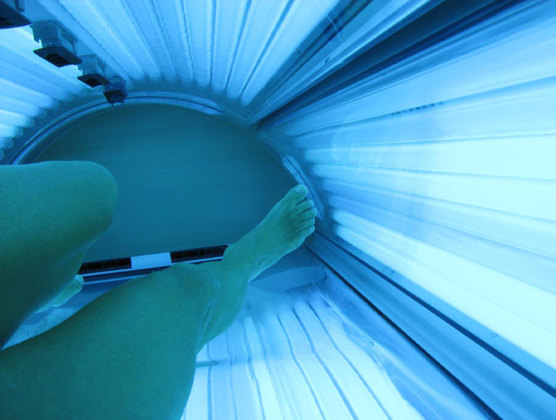
According to the American Academy of Dermatology and the U.S. Department of Health and Human Services, ultraviolet (UV) radiation from tanning beds, tanning booths and sun lamps are known carcinogens (cancer-causing substances). Exposure to UV radiation during indoor tanning has been proven to increase the risk of all skin cancers, including melanomas, squamous cell carcinomas and basal cell carcinomas. In fact, the risk of melanoma increases by 75 percent when indoor tanning devices are used before the age of 30. The UV radiation during indoor tanning also leads to skin aging, hyper – and hypopigmentation, immune suppression and eye damage, such as cataracts.

Therefore, the use of tanning beds, tanning booths and sun lamps is not recommended by dermatologists.

Persons legs inside tanning bed image

Psoriasis

Psoriasis is a skin condition that creates red patches of skin with white, flaky scales. It most commonly occurs on the elbows, knees and trunk, but can appear anywhere on the body. The first episode usually strikes between the ages of 15 and 35. It is a chronic condition that will then cycle through flare-ups and remissions throughout the rest of the patient’s life. Psoriasis affects as many as 7.5 million people in the United States. About 20,000 children under age 10 have been diagnosed with psoriasis.

In normal skin, skin cells live for about 28 days and then are shed from the outermost layer of the skin. With psoriasis, the immune system sends a faulty signal which speeds up the growth cycle of skin cells. Skin cells mature in a matter of 3 to 6 days. The pace is so rapid that the body is unable to shed the dead cells, and patches of raised red skin covered by scaly, white flakes form on the skin.

Psoriasis is a genetic disease (it runs in families), but is not contagious. There is no known cure or method of prevention. Treatment aims to minimize the symptoms and speed healing.

Skin Psoriasis Vs Eczema

Types of Psoriasis

There are five distinct types of psoriasis:

  • Plaque Psoriasis (Psoriasis Vulgaris)  About 80% of all psoriasis sufferers get this form of the disease. It is typically found on the elbows, knees, scalp and lower back. It classically appears as inflamed, red lesions covered by silvery-white scales.
  • Guttate Psoriasis This form of psoriasis appears as small red dot-like spots, usually on the trunk or limbs. It occurs most frequently among children and young adults. Guttate psoriasis comes on suddenly, often in response to some other health problem or environmental trigger, such as strep throat, tonsillitis, stress or injury to the skin.
  • Inverse Psoriasis This type of psoriasis appears as bright red lesions that are smooth and shiny. It is usually found in the armpits, groin, under the breasts and in skin folds around the genitals and buttocks.
  • Pustular Psoriasis  Pustular psoriasis looks like white blisters filled with pus surrounded by red skin. It can appear in a limited area of the skin or all over the body. The pus is made up of white blood cells and is not infectious. Triggers for pustular psoriasis include overexposure to ultraviolet radiation, irritating topical treatments, stress, infections and sudden withdrawal from systemic (treating the whole body) medications.
  • Erythrodermic Psoriasis One of the most inflamed forms of psoriasis, erythrodermic psoriasis looks like fiery, red skin covering large areas of the body that shed in white sheets instead of flakes. This form of psoriasis is usually very itchy and may cause some pain. Triggers for erythrodermic psoriasis include severe sunburn, infection, pneumonia, medications or abrupt withdrawal of systemic psoriasis treatment.

People who have psoriasis are at greater risk for contracting other health problems, such as heart disease, inflammatory bowel disease and diabetes. It has also been linked to a higher incidence of cardiovascular disease, hypertension, cancer, depression, obesity and other immune-related conditions.

Psoriasis triggers are specific to each person. Some common triggers include stress, injury to the skin, medication allergies, diet and weather.

Treatment

Psoriasis is classified as Mild to Moderate when it covers 3% to 10% of the body and Moderate to Severe when it covers more than 10% of the body. The severity of the disease impacts the choice of treatments.

Mild to Moderate Psoriasis

Mild to moderate psoriasis can generally be treated at home using a combination of three key strategies: over-the-counter medications, prescription topical treatments and light therapy/phototherapy.

Over-the-Counter Medications

The U.S. Food and Drug Administration has approved of two active ingredients for the treatment of psoriasis: salicylic acid, which works by causing the outer layer to shed, and coal tar, which slows the rapid growth of cells. Other over-the-counter treatments include:

  • Scale lifters that help loosen and remove scales so that medicine can reach the lesions.
  • Bath solutions, like oilated oatmeal, Epsom salts or Dead Sea salts that remove scaling and relieve itching.
  • Occlusion, in which areas where topical treatments have been applied are covered to improve absorption and effectiveness.
  • Anti-itch preparations, such as calamine lotion or hydrocortisone creams.
  • Moisturizers designed to keep the skin lubricated, reduce redness and itchiness and promote healing.

Prescription Topical Treatments

Prescription topicals focus on slowing down the growth of skin cells and reducing any inflammation. They include:

  • Anthralin, used to reduce the growth of skin cells associated with plaque.
  • Calcipotriene, that slows cell growth, flattens lesions and removes scales. It is also used to treat psoriasis of the scalp and nails.
  • Calcipotriene and Betamethasone Dipropionate. In addition to slowing down cell growth, flattening lesions and removing scales, this treatment helps reduce the itch and inflammation associated with psoriasis.
  • Calcitriol, an active form of vitamin D3 that helps control excessive skin cell production.
  • Tazarotene, a topical retinoid used to slow cell growth.
  • Topical steroids, the most commonly prescribed medication for treating psoriasis. Topical steroids fight inflammation and reduce the swelling and redness of lesions.

Light Therapy/Phototherapy

Controlled exposure of skin to ultraviolet light has been a successful treatment for some forms of psoriasis. Three primary light sources are used:

  • Sunshine (both UVA and UVB rays). Sunshine can help alleviate the symptoms of psoriasis, but must be used with careful monitoring to ensure that no other skin damage takes place. It is advised that exposure to sunshine be in controlled, short bursts.
  • Excimer lasers. These devices are used to target specific areas of psoriasis. The laser emits a high-intensity beam of UVB directly onto the psoriasis plaque. It generally takes between 4 and 10 treatments to see a tangible improvement.
  • Pulse dye lasers. Similar to the excimer laser, a pulse dye laser uses a different wavelength of UVB light. In addition to treating smaller areas of psoriasis, it destroys the blood vessels that contribute to the formation of lesions. It generally takes about 4 to 6 sessions to clear up a small area with a lesion.

Moderate to Severe Psoriasis

Treatments for moderate to severe psoriasis include prescription medications, biologics and light therapy/phototherapy.

Oral medications. This includes acitretin, cyclosporine and methotrexate. Your doctor will recommend the best oral medication based on the location, type and severity of your condition.

Biologics. A new classification of injectable drugs, biologics are designed to suppress the immune system. These tend to be very expensive and have many side effects, so they are generally reserved for the most severe cases.

Light Therapy/Phototherapy. Controlled exposure of skin to ultraviolet light has been a successful treatment for some forms of psoriasis. Two primary light sources are used:

  • Sunshine (both UVA and UVB rays). Sunshine can help alleviate the symptoms of psoriasis, but must be used with careful monitoring to ensure that no other skin damage takes place. It is advised that exposure to sunshine be limited to controlled, short bursts.
  • PUVA. This treatment combines a photosensitizing drug (psoralens) with UVA light exposure. This treatment takes several weeks to produce the desired result. In some severe cases, phototherapy using UVB light may lead to better results.

Diabetes-Related Skin Conditions

Diabetes and Its Impact on Skin Health

It is estimated that about one-third of people with diabetes will have a skin disorder at some time in their lives caused by the disease. Diabetics are more susceptible to bacterial and fungal infections; allergic reactions to medications, insect bites or foods; dry itchy skin as a result of poor blood circulation; and infections from foot injuries for people with neuropathy.

There are a number of diabetes-specific skin conditions:

Acanthosis Nigricans. A slowly progressing skin condition, which turns some areas of skin, usually in the folds or creases, into dark, thick and velvet-textured skin. Acanthosis nigricans often precedes the diagnosis of diabetes. It is sometimes inherited, but is usually triggered by high insulin levels. It can occur at any age and usually strikes people who are obese. There is no treatment for the condition except to reduce insulin levels. Prescription creams may help lighten the affected area.

Diabetic Blisters. Rare blisters that appear on the hands, toes, feet or forearms that are thought to be caused by diabetic neuropathy.

Diabetic Dermopathy. Round, brown or purple scaly patches that most frequently appear on the front of the legs (most often the shins) and look like age spots. They are caused by changes in small blood vessels. Diabetic dermopathy occurs more often in people who have suffered from diabetes for decades. They are harmless, requiring no medical intervention, but they are slow to heal.

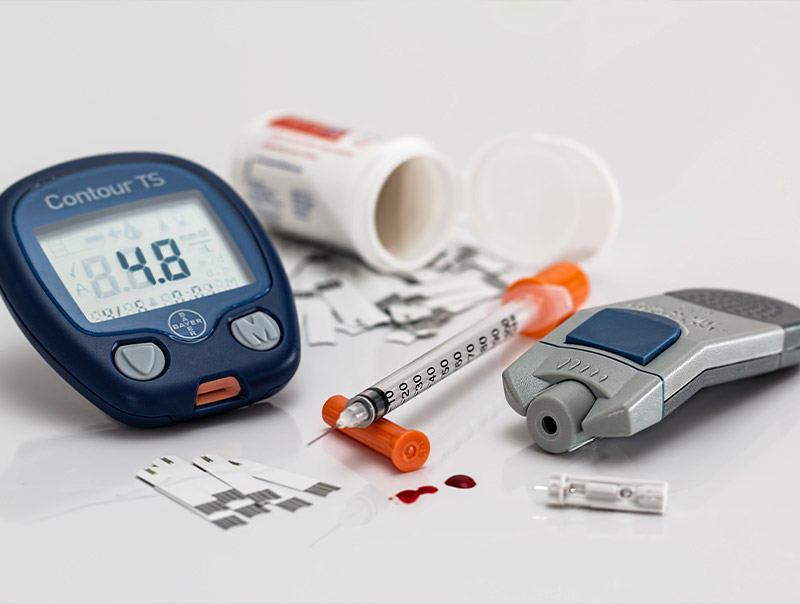
Various medical devices image

Digital Sclerosis. This condition appears as thick, waxy and tight skin on toes, fingers and hands, which can cause stiffness in the digits. Getting blood glucose levels back to normal helps alleviate this skin condition.

Disseminated Granuloma Annulare. A red or reddish-brown rash that forms a bull’s eye on the skin, usually on the fingers, toes or ears. While not serious, it is advised that you talk to your dermatologist about taking steroid medications to make the rash go away.

Eruptive Xanthomatosis. A pea-like enlargement in the skin with a red halo that itches. It most frequently appears on the hands, feet, arms, legs or buttocks. It is often a response to high triglycerides. Keeping blood glucose levels in the normal range helps this condition subside.

Necrobiosis Lipoidica Diabeticorum. This condition is similar to diabetic dermopathy, but the spots are larger, fewer, deeper in the skin and have a shiny porcelain-like appearance. It is often itchy or painful. It goes through cycles of being active and inactive. It is caused by changes in collagen and fat underneath the skin. Women are three times more likely to get this condition than are men. Typically, topical steroids are used to treat necrobiosis lipoidica diabeticorum. In more severe cases, cortisone injections may be required.

Vitiligo. Vitiligo refers to the development of white patches anywhere on the skin. It usually affects areas of skin that have been exposed to sun. It also appears in body folds, near moles or at the site of previous skin injury. The condition is permanent and there is no known cure or prevention. However, there are some treatments that can be used to improve the appearance of the skin, such as steroid creams and ultraviolet light therapy.

Pregnancy-Related Skin Conditions

Although less common, there are a few skin conditions related to pregnancy:

PUPPP (Pruritic Uticarial Papules and Plaques of Pregnancy)

This condition occurs in roughly one percent of pregnant women. It is characterized by itchy red bumps and hive-like rashes that usually appear on the belly or around stretch marks. The rash may spread to the arms, legs, breasts or buttocks. PUPPP usually begins in the third trimester of pregnancy. It is harmless, but the itchiness can be severe. There is no known cause for the condition. Treatment typically includes the use of topical ointments, antihistamines and, in more severe cases, oral steroids to help alleviate the itch. PUPPP usually disappears a few days after the baby’s birth.

Pregnant woman with ocean in background image

Prurigo of Pregnancy (Papular Eruptions of Pregnancy)

A rare skin condition that can occur anywhere on the body. Prurigo looks like a collection of bug bites. Its onset is usually not before the third trimester and it typically lasts up to three months after delivery. The condition is harmless to mother and baby. Like PUPPP, it is generally treated with topical ointments, oral medications, antihistamines or steroids.

Pemphigold Gestationis (Herpes Gestationis)

This extremely rare condition starts as a hive-like rash, which turns into large blistering lesions. It usually begins on the abdomen and spreads to the mother’s arms and legs. It causes severe itchiness. It typically begins in either the second or third trimester. It may also come and go intermittently throughout a pregnancy. Pemphigold gestationis is associated with an increased risk for pre-term delivery and fetal health issues. If you suspect you may have this condition, seek immediate medical attention.

Our Info

Contact